-
For patient reports Click Here
For patient reports Click Here


Over the years, Tesla Diagnostics has been at the epicenter for providing care and services in the field of cardiology. Today, cardiovascular diseases are assumed to be the leading cause of death. Cholesterol, high blood pressure, family and genetic history, unhealthy lifestyle are some of the leading causes of cardiovascular diseases. Prevention may go a long way in combating heart diseases. A series of cardiovascular tests are available to determine the cause of irregular heartbeats, unexplained chest pain, dizziness, shortness of breath, etc.
In some cases, patients with coronary blockages may have normal E.C. G. ECGs are taken at rest but symptoms of heart disease may become unmasked when the heart is exposed to stress or exercise. Under the stress of exercise, the healthy coronary arteries dilate or open up more than that of an artery with blockage. This results in more delivery of blood to the heart muscles. On the other hand, blocked arteries result in less delivery of blood causing the muscles to “starve”. This may lead to uneasiness like chest discomfort, shortness of breath which an ECG fails to detect at rest.
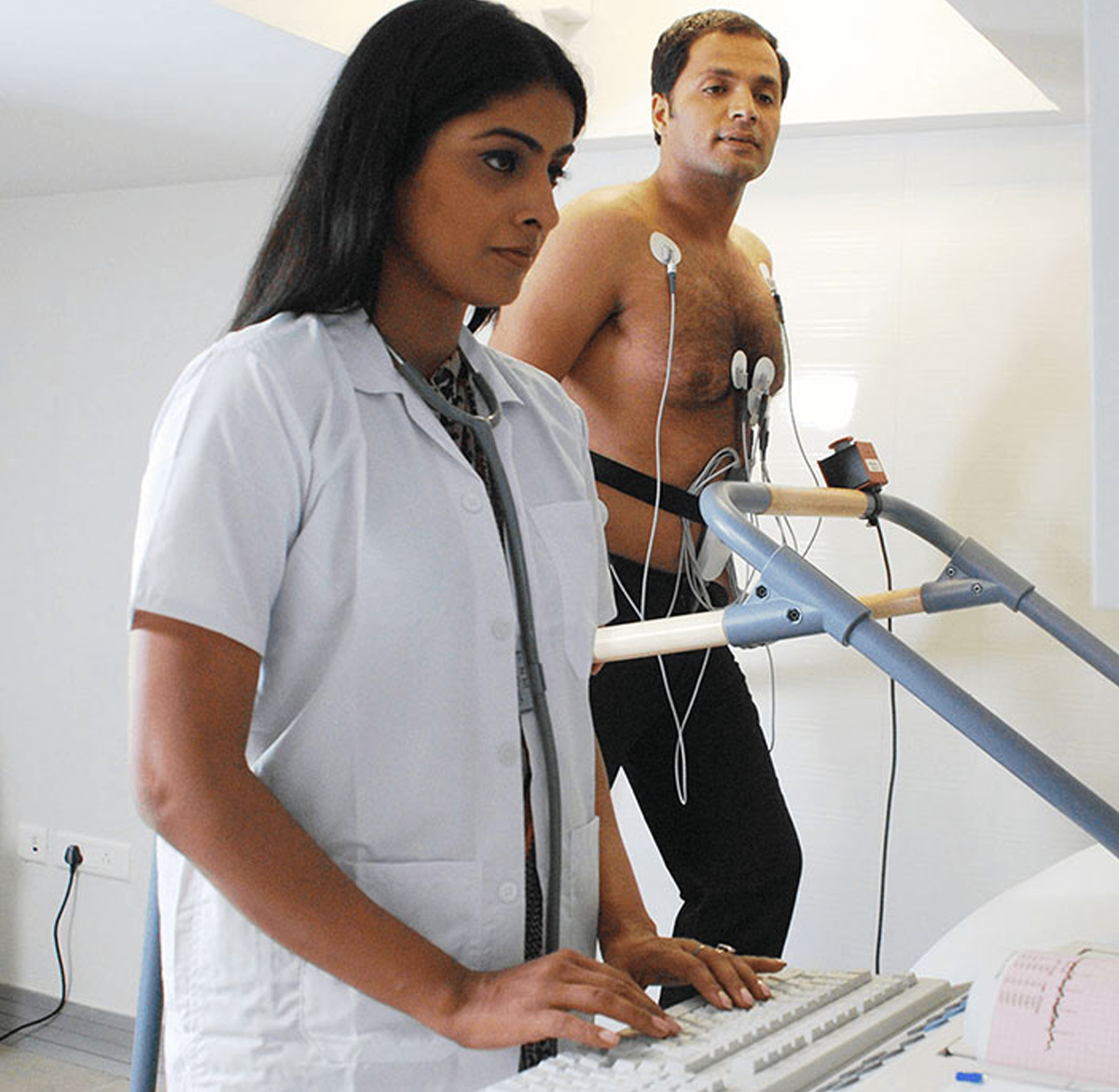
At Tesla, the leading cardiology hospital, we perform the TMT or Treadmill Test to record the stress on the heart. It is a non-invasive test in which the patient is made to walk on a treadmill. The heart rate and blood pressure of the patient at rest are noted. Then sticky electrodes are attached to the chest, shoulders and hips to monitor the heart with the help of an ECG and the blood pressure is also monitored at regular intervals. The patient is made to walk on the treadmill and the speed is gradually increased according to a preprogrammed protocol until the target heart rate is reached. The 12-lead ECG is recorded on paper. The heart rate, changes in the ECG pattern, blood pressure, and irregular heart rhythm is evaluated. The test is terminated prior to achieving the target rate in case the ECG shows changes or the patient experiences any discomfort or chest pain or breathlessness. It may also be stopped if the BP falls above or below the acceptable limits.
If you show signs or symptoms of a coronary artery disease
If you have significant risk factors for a coronary artery disease
If you have unexplained fatigue or shortness of breath
To check your blood pressure as an effect of exercise in case you have borderline hypertension
To screen for irregular heartbeats induced by stress
At Tesla, the test is performed using a high-end GE machine by qualified and experienced technicians, under the supervision of experienced physicians and cardiologists.
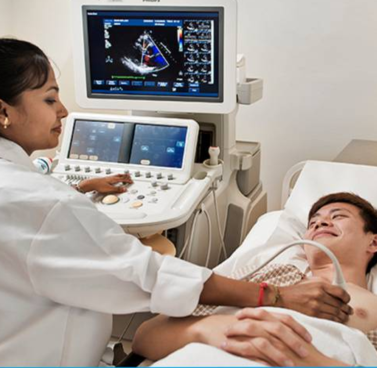
2D Echo or Echocardiogram or cardiac ultrasound or simply an ‘echo’ is a non-invasive, imaging technique used to get a clear picture of the heart. It displays a cross-sectional slice of the heart including the valves, chambers and blood vessels that enter and exit from the heart. It is used to assess the functions and structures of the heart. A transducer (similar to a microphone) is used in the process that emits ultrasonic sound waves at a very high frequency such that it cannot be heard by the human ear. This transducer is placed on the chest of the patient at specific angles and locations, such that the ultrasonic waves pass through the skin and body tissues to reach the heart. The waves bounce off the heart structures and are sent to a computer to create moving images of the heart structures.
Generally, no preparation is required for the test. It is recommended to inform the doctor about any medications you are taking or if you have a pacemaker installed. A colorless gel is applied on the chest and /or on the transducer head. The room would be darkened so that images can be viewed clearly. The transducer probe is placed on different parts of your chest to get views of specific regions of the heart. The resultant images are viewed on a monitor and recorded on photographic paper; computer discs or videotape and the readings are reviewed and interpreted by the cardiologist.
Your cardiologist or physician may suggest an echo for the following reasons:
To detect heart chamber abnormalities such as those with size and dimensions
To detect abnormalities in the wall motion or pumping of the heart
To detect congenital heart diseases.
To detect any conditions that affect the heart muscles, valves or pericardium,
To detect abnormalities of the heart valves pertaining to the structure, thickness or movement
To evaluate heart murmurs
The test results provide your cardiologist with information pertaining to the structure and functioning of the heart and blood flow. This helps them to plan their line of treatment. At Tesla, we are equipped with the latest, Philips HD 11 machine with specialized Mindray cardiac probes.
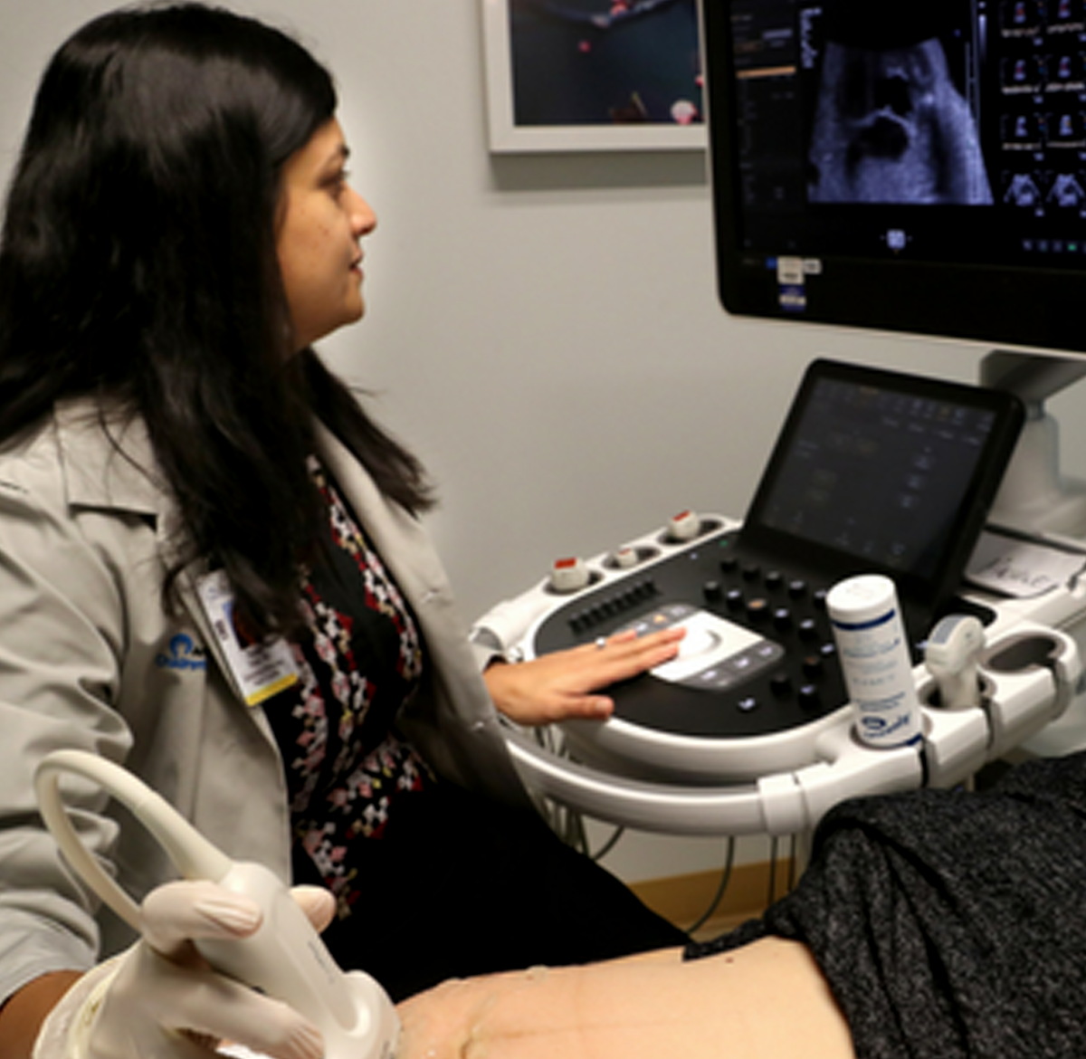
Fetal Echocardiography is a diagnostic test done while the baby is still in the womb. It is a detailed scan of the heart to detect heart defects. It is usually performed in the second trimester i.e. between 18-24 weeks of pregnancy. It helps the doctor to get a clear picture of the structure and functions of the fetal heart. It is very similar to that of ultrasound and uses sound waves that bounce off the surface of the heart’s structures. These sound waves are analyzed and a picture of the baby’s heart is created on the computer which provides information about the structure of the heart. Additionally, it also allows viewing the blood flow through the heart and detects anomalies in heartbeat or blood flow if any.
The fetal echo test provides a better picture compared to regular pregnancy ultrasound. It shows
Blood flow through the fetal heart
The rhythm of the fetal heart
Structures of the baby’s heart
Your doctor may prescribe this test if
There is a family history of heart disease/defect
You already have a child with a heart defect/disease
If the mother has medical conditions like rubella, lupus, type 1 diabetes, phenylketonuria
If the mother has used drugs or alcohol during pregnancy
If the mother has taken medicines such as those for epilepsy
The test can either be done on your abdomen known as an abdominal ultrasound or through the vagina known as transvaginal ultrasound.
An abdominal ultrasound involves placing a gel on your abdomen and moving a handheld probe over the area. The sound waves from the probe reach the baby’s heart and bounce off creating a picture of your baby’s heart on a computer.
Transvaginal ultrasound involves inserting a smaller probe into the vagina. This provides a clearer image when compared to abdominal ultrasound and can be done earlier in pregnancy. There are no associated risks to the mother or the unborn baby. If a problem is detected, a detailed ultrasound may be recommended. Tesla Diagnostics is equipped with a high-end Philips HD 11 machine and the procedure is performed in the presence of a highly experienced team of cardiologists.
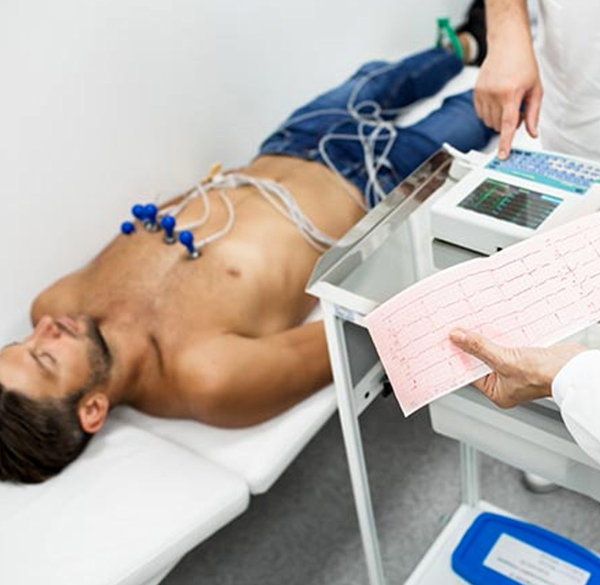
An ECG or an Electrocardiogram or EKG is a simple, non-invasive, painless test that records the heart’s electrical activity and helps detect any abnormalities in the heart. Let us understand how the heart works in order to understand the test.
An electrical signal spreads from the top of the heart to the bottom during each heartbeat. As the signal passes through the heart it causes the heart to contract and pumps out blood to the lungs and rest of the body. These signals set the heartbeat rhythm.
These electrical signals are translated as line tracings on paper by an ECG. ECG from normal healthy people has a characteristic shape whereas those from people with heart problems show a change in shape as there is a change in the electrical activity of their heart.
Your doctor may recommend an ECG test if you are at risk of heart diseases because of a family history of high cholesterol or high blood pressure or smoking or obesity or the like. An ECG helps detect the following problems of the heart
Enlargement of the heart
Congenital defects that involve the electrical system of the heart
The irregular rhythm of the heart also known as arrhythmia
Blocked or narrowed arteries known as coronary occlusion
Inflammation in the heart (pericarditis or myocarditis)
Heart Attacks during intensive care monitoring or in emergency rooms
Abnormality with Heart valves
An ECG can also help
To find medical conditions that cause chest pain, shortness of breath, fainting, dizziness, palpitations (irregular heartbeats)
To assess the condition of people diagnosed with heart disease
To assess the functioning of devices such as pacemakers within the body
To check the effect and side effects of certain medications on the heart.
Before performing an ECG, inform your doctor of any medicines that you may be taking as certain medicines influence the test results. Electrodes from an ECG machine are attached to the skin of the arms, chest and legs using sticky patches or suction cups. These electrodes read signals from the heart and send it to the ECG machine. The machine then generates the results as a series of spikes and dips which is interpreted by a cardiologist. There are three types of ECG
Resting ECG- is performed in a lying position. No movement is allowed and it takes only 10-15 minutes
Ambulatory ECG or 24 hour ECG- is performed using a small electrocardiograph machine worn throughout the day. The patient may move around while the device is attached. It is used in patients who show intermittent symptoms that are not visible during a resting ECG.
Cardiac Stress Test or Exercise ECG- is done while the patient is exercising usually walking on a treadmill. It takes approximately 15-30 minutes to complete.
There are rarely any risks associated with the test. It helps the cardiologist to detect abnormalities in the heart and plan their line of treatment. If you are searching for a cardiologist near me or an ECG test near me, then reach Tesla which is equipped with a state of the art equipment and experienced doctors and technicians who carry out the test.